ISSN 2954-467X

Inconclusive prenatal sex determination in an NIPT due to probable confined placental mosaicism and the importance of amniocentesis.


Ronny Kershenovich Sefchovich


Ronny Kershenovich Sefchovich1, 0009-0000-2539-3013; Héctor Oviedo Cruz2, 0000-0001-8781-5592; Marcela Fragoso Benitez3, 0000-0002-4218-9951; Leonardo Pérez Mejía4, 0009-0001-5797-8857; Rolando Álvarez Valero1, 0009-0002-4790-1634.


1 American British Cowdray Medical Center (Centro Médico ABC).
2 Centro Médico Para Atención Fetal Especializada (CEMAFE).
3 GD Technologies.
4 Genos Médica.

NOTE: The numbers following the affiliation markers are the author's ORCID iD.

Contact: Dr. Ronny Kershenovich Sefcho. mail: genetista@me.com. American British Cowdray Medical Center, Av. Carlos Graef Fernández, 154, Colonia Santa Fe, Cuajimalpa, CP 05300, Mexico City, CEGOP Building, Lobby, Office 1B. Phone: +52-5516647227.

Article History: Received July 11th, 2023. Revised July 21th, 2023. Accepted July 25th, 2023. Available online August 29th, 2023.


Abstract

Non-invasive prenatal diagnosis in maternal blood from placental has become the first-line test for the early detection of chromosomal aneuploidies. Amniocentesis remains the gold standard for the accurate diagnosis of any chromosomal aneuploidy. However, sometimes noninvasive prenatal tests can report inconclusive results which presents a dilemma for decision making. We report a case where fetal sex was inconclusive and confirmation by amniocentesis was performed on a couple.


Keywords

NIPT, amniocentesis, sex determination, mosaicism, inconclusive.


Manuscript


Introduction


Non-invasive prenatal diagnosis (NIPT) has revolutionized the way prenatal screening can be done in an early, quick, and reliable manner since its first clinical use in 2011. Since its adoption in 2016 for use in all women regardless of age by the American College of Medical Genetics and Genomics, its use has been increasing worldwide as a first-tier method for the detection of chromosomal aneuploidies such as trisomies 13, 18 and 21 and sex chromosome aneuploidies such as monosomy X, XXX, XXY and XYY (1). Importantly, pre-test and post-test genetic counseling should be offered to all couples regardless of their reproductive history. However, as NIPT involves the use of cells originating from the placenta in the maternal bloodstream there is the possibility of inconclusive results due to low fetal fraction or mosaicism. We must remember that the placenta is an organ generated exclusively during pregnancy for the blood supply and nutrients from mother to the fetus and vice versa. That said, placental cells will not always have the same ploidy of chromosomes as the fetus, and this can lead to this type of results due to the presence of probable confined placental mosaicism (CPM), fetal confined mosaicism (FCM) or mixed mosaicism (2,3) in up to 2% of all pregnancies (Figure 1). CPM is usually identified after the first trimester with invasive chorionic villus diagnosis and when mosaicism is detected, amniocentesis is performed to determine if the abnormal cell line is also present in fetal tissues. Amniotic fluid karyotype is usually reported as normal in most cases (72% to 87%) (4,5) In addition, CPM can also be found in cytogenetic studies of the placenta of euploid fetuses (6-8) and is recognized as a relevant source of false-positive results in NIPT (9,10). CPM can be classified into three subtypes (type 1, 2, and 3) depending on where the chromosomal abnormality is in the placenta (Fig. 1) (Toutain et al., 2018). When the chromosomal abnormality is only found in the cytotrophoblast (and can be found after short-term culture villus examination (STC), it is CPM type 1. If the chromosomal abnormality is only found after a long-term villi culture (LTC), it is restricted to the mesenchymal nucleus of chorionic villi and is classified as type 2. Type 3 is defined as the presence of the abnormality in both the mesenchymal nucleus and the cytotrophoblast and can be found after LTC and STC analysis. As mentioned above, NIPT (and STC-villi) analyzes cytotrophoblast and therefore, NIPT can determine CPM type I and type III (11).


figure 1

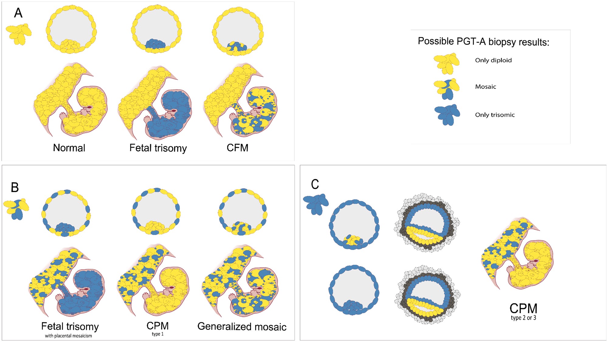
Figure 1. From blastocyst cells to prenatal scenario. (A) Three different scenarios arise if only diploid cells are retrieved through biopsy in blastocyst stage. (B) When both diploid and trisomic cells are biopsied, three different scenarios can also arise. If the mosaicism is only found in the trophectoderm and not within the inner cell mass (ICM), confined placental mosaicim (CPM) type 1 develops. (C) If all biopsied cells appear to be trisomic, even in the ICM, as a result of trisomic rescue, the epiblast will eliminate the trisomic cells and will only consist of diploid cells, thus CPM type 2 or 3 can develop.Taken from Toutain et al., 2018.

Figure use license: https://doi.org/10.1093/humupd/dmab009


Case presentation


We present a case of a couple Gestation 2 Abortion 1 where both are already of advanced age (38 and 46 years respectively) where there was a previous molar pregnancy. They went to the medical geneticist for advice of a second pregnancy of 14.5 gestational weeks by in vitro fertilization (IVF) and in which they did not want to perform preimplantation genetic diagnosis for aneuploidies (PGT-A), only a NIPT was practiced at week 10.3 of pregnancy where they did not report aneuploidies, nor CNV's of the autosomal and sexual chromosomes, however, fetal sex was reported as "Fetal chromosomal sex not defined by the presence of probable Y chromosome mosaicism" (Figure 2). They were provided with genetic counseling in the first instance and opted for amniocentesis to rule out the presence of mosaicism through a karyotype. Mosaicism at this stage is important to understand that it can be confined to placenta, confined to the fetus or mixed (Figure 1) where the result can have important consequences for the fetus and the course of pregnancy if no reported anomaly is verified, so amniocentesis is always necessary and that is why it still remains as the gold standard for a diagnostic method. Amniocentesis in the mother and fetus were performed by a maternal-fetal physician and sent for analysis. The karyotype result was reported as 46,XY [30], which corresponds to a male fetus in 30 metaphases (Figure 2).


figure 2a

(a)

figure 2b

(b)


Discussion


Mosaicism is an anomaly that can occur in up to 2% of cases. It is important to note that they can be of 3 types depending on their embryological origin, thus giving 3 types, the CPM, FCM and generalized mosaicism. Regardless of whether it is a PCM, FCM or a generalized mosaicism, it can lead to serious problems in a pregnancy either spontaneously or by assisted reproduction techniques such as aneuploidies that are not compatible with life. In this case report, we demonstrate the importance of performing amniocentesis following a noninvasive prenatal test on maternal blood where fetal sex was reported as inconclusive. Amniocentesis is still the gold standard for determining with 100% accuracy the presence or absence of aneuploidies of autosomes and sex chromosomes when reported as inconclusive by probable mosaicism. In our case it was determined that it was a mosaicism confined to the placenta since only a single cell line was found in the fetal cells of the amniotic fluid.


Conclusions


Before a pregnancy performed by IVF, it is advisable to perform PGT-A as it can help drastically to make decisions. When performing a non-invasive prenatal test on maternal blood that is inconclusive, it is imperative to perform amniocentesis to rule out or verify the presence of mosaicism.


Funding

This research did not receive any specific grant from funding agencies in the public, commercial, or not-for-profit sectors.


Conflict of interest

The authors declare that there is no conflict of interest.


References

[1] Gregg AR, Skotko BG, Benkendorf JL, et al. Noninvasive prenatal screening for fetal aneuploidy, 2016 update: a position statement of the American College of Medical Genetics and Genomics. Genet Med. 2016;18(10):1056-1065. http://doi.org/10.1038/gim.2016.97.

[2] Kalousek DK, Dill FJ. Chromosomal mosaicism confined to the placenta in human conceptions. Science 1983;221:665–7.

[3] Simoni G, Sirchia SM. Confined placental mosaicism. Prenat Diagn 1994;14:1185–9.

[4] Grati FR, Malvestiti F, Branca L, et al. Chromosomal mosaicism in the fetoplacental unit best practice & research. Clin Obstet Gynaecol 2017;42:39–52.

[5] Battaglia P, Baroncini A, Mattarozzi A, et al. Cytogenetic follow-up of chromosomal mosaicism detected in first-trimester prenatal diagnosis. Prenat Diagn 2014;34:739–47.

[6] Wilkins-Haug L, Quade B, Morton CC. Confined placental mosaicism as a risk factor among newborns with fetal growth restriction. Prenat Diagn 2006;26:428–32.

[7] Stipoljev F, Latin V, Kos M, Miskovic B, Kurjak A. Correlation of confined placental mosaicism with fetal intrauterine growth retardation. A case control study of placentas at delivery. Fetal Diagn Ther 2001;16:4–9.

[8] Kalousek DK, Howard-Peebles PN, Olson SB, et al. Confirmation of CVS mosaicism in term placentae and high frequency of intrauterine growth retardation association with confined placental mosaicism. Prenat Diagn 1991;11:743–50.

[9] Mardy A, Wapner RJ. Confined placental mosaicism and its impact on confirmation of NIPT results. Am J Med Genet C Semin Med Genet 2016;172:118–22.

[10] Brison N, Neofytou M, Dehaspe L, et al. Predicting fetoplacental chromosomal mosaicism during non-invasive prenatal testing. Prenat Diagn 2018;38:258–66.

[11] Toutain J, Goutte-Gattat D, Horovitz J, Saura R. Confined placental mosaicism revisited: Impact on pregnancy characteristics and outcome. PLoS One 2018;13:e0195905.